الگوریتم نرمال سازی شده تقسیم بندی تصاویر اتسو (Otsu) برای تشخیص بیماری ملانوما
چکیده
ملانوما یک سرطان پوستی کشنده است که میزان مرگ را با آهنگ سریع تری افزایش می دهد.برای اینکه این میزان مرگ کنترل شود بایستی ملانوما در مرحله ی اولیه ی آن کشف شود.برای دستیابی به این مهم محققان تشخیص به کمک کامپیوتر را معرفی کرده و این روش را برگزیده اند.الگوریتم های فراوانی برای چند تکه ای کردن تصویر وجود دارد که یکی از مهم ترین این الگوریتم ها ، الگوریتم مرسوم چند تکه ای اتسو می باشد. ایراد اصلی این الگوریتم این است که در حضور نورپردازی متغیر روش چند تکه ای مناسب نمی باشد. برای حل این نقیصه این مقاله الگوریتم چند تکه ای نرمال شده ی اتسو را پیشنهاد می کند که بر ایراد اشاره شده در فوق غلبه می کند و منجر به یک تقسیم بندی دقیق تصویر می شود. این الگوریتم ابتدا ، تصویر را نرمال سازی می کند تا بر نورپردازی متغیر غلبه کند و سپس با استفاده از الگوریتم اتسو ، تصویر را تکه تکه و چند قسمتی می کند.نتیجه ی دقیقی که این الگوریتم ارائه می کند می تواند در مراحل بیشتری مورد استفاده قرار گیرد تا ایراد رابطور دقیق کشف کند و در نتیجه به کاهش میزان مرگ کمک خواهد کرد.
مقدمه
بیماری ملانوما می تواند در مرحله ی اولیه با استفاده از تحلیل های کامپیوتری کمکی کشف شود که در این راستا چند تکه سازی تصویر گامی مهم و اساسی محسوب می شود. چند تکه سازی تصویر به کمک الگوریتم چند تکه ای انجام می شود تا سرعت پردازش را افزایش دهد.بعد از تقسیم تصویر به نواحی متعدد، اندازه گیری و سنجش روی هر ناحیه می تواند صورت گیرد.بنابراین چند تکه سازی گامی اساسی در آنالیز داده های تصویرمحسوب می شود. روش های متعددی برای چند تکه سازی پیشنهاد و ارائه شده است ناپیوستگی و همگن بودن دو شاخصه ای هستند که تعیین کننده ی نوع روش چند تکه سازی تصویر می باشند.تقسیم بندی ناحیه محور و تقسیم بندی لبه محور دو نمونه از انواع دسته بندی چندتکه سازی تصویر هستند که بر مبنای دو شاخص ذکر شده در فوق می باشند. بر اساس خاصیت ناپیوستگی پیکسل های تصویر، روش های چند تکه کردن تصویر بعنوان لبه ای یا مرزی طبقه بندی می شوند. تصویر مضاعف و دوتایی نتیجه ی تقسیم بندی لبه محور می باشد. دو نمونه از دسته بندی های روش چند تکه سازی لبه محور، روش های زاویه محور و هیستوگرام خاکستری می باشند.
6)نتیجه گیری
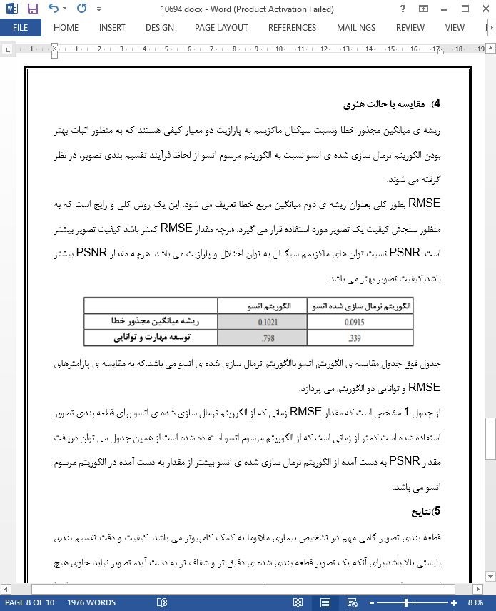
این مقاله الگوریتمی را ارائه می کند که می تواند به منظور به دست آمدن نتایجی دقیق تر و بهتر در فرآیند قطعه بندی تصویر ،به سادگی مورد استفاده قرار گیرد. و یک مسیر وسیع تر و بهتر را جهت پیش بینی بیماری ملانوما فراهم می کند.الگوریتم پیشنهاد شده در این مقاله میزان خطای کمتر و مقدار PSNR بیشتری را تولید می کند که ثابت می کند کیفیت تصویر تقسیم بندی شده بالا می باشد.زمانی که این الگوریتم برای تقسیم بندی تصویر مورد استفاده قرار می گیرد مشکل نورپردازی متغیر رفع می شود. زمانی که مشکل نورپردازی متغیر برطرف شود، نتیجه ی فرآیند تقسیم بندی تصویر دقیق تر و شفاف تر خواهد بود که یک روش گسترده تر و شفاف تر برای استخراج ویژگی ها و مشخصات از تصویر قطعه بندی شده را فراهم می کند به نحوی که تصویر دارای کیفیت بالاتری می باشد.
Abstract
Melanoma is a deadly skin cancer which increases the death rate at a faster rate. In order to bring the death rate under control, melanoma should be detected at its earlier stage. To achieve this, researchers have introduced Computer aided diagnosis and adopted the same. In this technique, Segmentation is found to be one of the important steps. Many algorithms exist in practise for segmentation,where one of the important algorithms is Traditional Otsu Segmentation Algorithm. In this algorithm the major drawback is that the segmentation is improper in the presence of variable illumination. This paper proposes an algorithm “Normalised Otsu Segmentation” which overcomes the above mentioned drawback and results in an accurate segmentation. This algorithm first normalises the image to overcome variable illumination and then segments the image using Otsu algorithm. The accurate result given by this algorithm can be used in further steps to detect the lesion accurately which will provide a hand a for reducing the death rate.
1. Introduction
Melanomacan be detected at earlier stage using computer aided analysis in which segmentation is the major and important step1-3. Segmentation is done with the help of Segmentation algorithm to speed up the process. After segmenting the image into subregions measurements on each region can be accomplished4 . Therefore segmentation is a major step for significant analysis of image data. Various techniques have been proposed for segmentation in many literatures. Discontinuity and similarity are the two properties based on which categorization of image segmentation method is done5 . Region based and edge based segmentation are the two categories of image segmentation based on these properties. Based on the property of discontinuity of pixels segmentation methods are classified as edge or boundary based techniques. A binary image is the result of edge based segmentation. The two categories of edge based segmentation methods are gradient based and gray histogram based methods6 .
6. Conclusion
This literature provides an algorithm which can be implemented easily for obtaining better and accurate results in the segmentation process, which provides a better and wider path for melanoma prediction. The proposed algorithm of this literature produces less error rate and high PSNR value which proves that the segmented image quality is high. When this proposed algorithm is used for segmentation, the problem of variable illumination can be overcome. When this variable illumination problem is removed,the result of the segmentation process is more accurate and clear, which provides a wider and clear way for extracting features from the segmented image in more qualified and quantified manner.

چکیده
1) مقدمه
2) الگوریتم پیشنهادی
3) اجرای الگوریتم
4) مقایسه با حالت هنری
5) نتایج
6) نتیجه گیری
Abstract
1. Introduction
2 Proposed Algorithm
3. Implementation
4. Comparison with State of Art
5. Results
6. Conclusion
- اصل مقاله انگلیسی با فرمت ورد (word) با قابلیت ویرایش
- ترجمه فارسی مقاله با فرمت ورد (word) با قابلیت ویرایش، بدون آرم سایت ای ترجمه
- ترجمه فارسی مقاله با فرمت pdf، بدون آرم سایت ای ترجمه
