تشخیص خودکار و اندازه گیری فضای شفاف پشت گردن جنین (نوکال ترانس لوسنسی)
چکیده
در این مقاله، ما یک روش شناسی جدید را برای حمایت از پزشکان به منظور شناسایی خودکار منطقه ی پشت گردن و برای دستیابی به اندازه گیری صحیح ضخامت فضای شفاف پشت گردن پیشنهاد کردیم. ضخامت فضای شفاف پشت گردن، یکی از مارکرهای اصلی غربال گری نقایص کروموزومی مانند تریزومی 13، 18 و 21 است. اندازه گیری آن در طی اسکن سونوگرافی در سه ماهه ی اول بارداری انجام می شود. روش شناسی پیشنهاد شده، اساسا بر اساس آنالیز موجک (ویولت) و تحلیل چند مقیاسی است. کارایی روش ما روی 382 فریم تصادفی که نشان دهنده ی برش (مقطع) میدساژیتال بودند و به صورت یکنواخت از روی فیلم های سونوگرافی بالینی واقعی 12 بیمار به دست آمده بودند، آنالیز شد. مطابق با پاسخ های صحیح مبنا ارائه شده توسط یک پزشک متخصص، ما یک نرخ مثبت واقعی برابر 99.95% را با توجه به تشخیص منطقه ی پشت گردن به دست آوردیم و حدود 64% از اندازه گیری ها، یک خطای برابر 1 پیکسل (که متناظر با 0.1 میلی متر است) داشتند.
1. مقدمه
سندرم داون (یا DS)، در سال 1886 توسط دکتر لانگدان داون شناسایی شد، این سندرم، یک بیماری ژنتیکی است که موجب ایجاد درجات متفاوتی از عقب ماندگی ذهنی، نمو جسمی و حرکتی می شود. آن توسط حضور یک کروموزوم اضافی در جفت 21ام در هسته های هر سلول (47 تا در مقایسه با تعداد نرمال که برابر 46 است) مشخص می شود؛ به این دلیل، به DS اغلب تحت عنوان تریزومی 21 نیز اشاره می شود. دلایل آن هنوز مشخص نیست و بنابراین هیچ روش واقعی برای پیش گیری از آن وجود ندارد. در اوایل دهه ی 70، سن مادر، اولین عاملی بود که مشخص شد ممکن است موجب ابتلا جنین به نقص کروزومومی شود.
5. نتایج
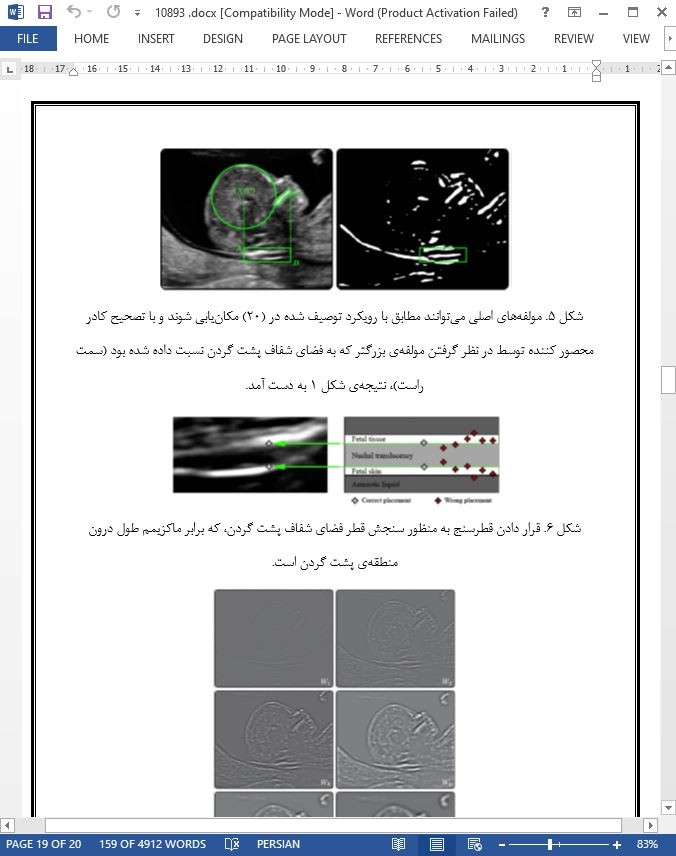
دستاورد پیشنهاد شده، یک روش شناسی خودکار را برای حمایت از پزشکان در ارزیابی برخی از نقایص کروموزومی مهم معرفی می کند. باید تاکید شود که، مطابق با پروتوکل بین المللی توصیف شده در (6)، فقط برش های (مقاطع) میدساژیتال باید در نظر گرفته شوند. در واقع، روش شناسی ما روی انتخاب این فریم ها از روی ویدئوها تکیه دارد که به دلیل نوع الگوریتم ماست که قبلا در (20) توصیف شده بود. اینجا ما توجه خودمان را روی تشخیص و اندازه گیری فضای شفاف پشت گردن متمرکز کرده ایم و از این رو یک محیط کامل را برای نشان دادن بیماری های احتمالی مانند تریزومی 13،18 و 21 در طی غربال گری سونوگرافی در طی سه ماهه ی اول بارداری ارائه کرده ایم. ما به صورت آزمایشگاهی، صحت روش شناسی را روی 382 فریم تصادفی به دست آمده از 12 معاینه ی واقعی تایید کردیم و نتایج به دست آمده را با پاسخ های صحیح مبنا ارائه شده توسط یک پزشک متخصص مورد مقایسه قرار دادیم.
Abstract
In this paper we propose a new methodology to support the physician both to identify automatically the nuchal region and to obtain a correct thickness measurement of the nuchal translucency. The thickness of the nuchal translucency is one of the main markers for screening of chromosomal defects such as trisomy 13, 18 and 21. Its measurement is performed during ultrasound scanning in the first trimester of pregnancy. The proposed methodology is mainly based on wavelet and multi resolution analysis. The performance of our method was analysed on 382 random frames, representing mid-sagittal sections, uniformly extracted from real clinical ultrasound videos of 12 patients. According to the ground-truth provided by an expert physician, we obtained a true positive rate equal to 99.95% with respect to the nuchal region detection and about 64% of measurements present an error equal to 1 pixel (which corresponds to 0.1 mm), respectively.
1. Introduction
Down's Syndrome (namely DS), identified in 1886 by Dr. Langdon Down, is a genetic condition that causes a variable degree of delay in mental, physical and motor development. It is caused by the presence of an extra chromosome in the nucleus of every cell (47 in comparison with a normal number of 46) in the twenty-first pair; for this reason DS is often indicated as trisomy 21. Its causes are still unknown and therefore there is no real way of prevention. Early in the 70's, the maternal age was the first element to deduce the probability for the fetus to present a chromosomal defect.
5. Conclusions
The proposed contribution introduces an automatic methodology to support physicians in the evaluation of some important chromosomal defects. It must be stressed that, according to the international acquisition protocol described in [6], only mid-sagittal sections must be considered. Indeed, our methodology relies on the selection of those frames from video sequences, due to our algorithm already described in [20]. Here we focus our attention on detecting and measuring the nuchal translucency, thus presenting a complete environment to highlight eventual diseases like trisomy 13, 18 and 21 during ultrasound scanning in the first trimester of pregnancy. We experimentally verified the correctness of our methodology on 382 random frames from 12 real examinations and compared the obtained results against the groundtruth provided by an expert physician.

چکیده
1. مقدمه
2. کارهای مرتبط
3. مواد و روش ها
3.1. توصیف مجموعه داده ها
3.2. پردازش تصویر موجک
3.3. تشخیص کادر محصور کننده ی پشت گردن
3.4. اندازه گیری منطقه ی شفاف پشت گردن
4. نتایج و بحث ها
4.1. انتخاب آستانه ی مناسب
4.2. ارزیابی ضخامت و منطقه ی پشت گردن
5. نتایج
ABSTRACT
1. Introduction
2. Related work
3. Materials and methods
3.1. Dataset description
3.2. Wavelet image processing
3.3. Nuchal bounding box detection
3.4. Measurement of the nuchal translucency
4. Results and discussions
4.1. Proper threshold selection
4.2. Nuchal region and thickness evaluation
5. Conclusions
- اصل مقاله انگلیسی با فرمت ورد (word) با قابلیت ویرایش
- ترجمه فارسی مقاله با فرمت ورد (word) با قابلیت ویرایش، بدون آرم سایت ای ترجمه
- ترجمه فارسی مقاله با فرمت pdf، بدون آرم سایت ای ترجمه
