دیستروفی اپیدرمولیز بولوزا: یک مرور
چکیده
دیستروفی اپیدرمولایزیس بولوسا یک اختلال نادر توارثی است که بر اثر جهش در ژن COL7A1 کد کننده ی کلاژن تیپ VII به وجود می آید. ناکارآمدی و/یا اختلال کلاژن تیپ VII باعث ایجاد تاول های زیرپوستی دقیقا زیر لامینا دنسا می شود که منجر به شکنندگی پوستی مخاطی و پیچیدگی بیماری مانند زخم های خودسر، خراش های گسترده، سوءتغذیه و بدخیمی می گردد. این بیماری ها اغلب با نقشه برداری ایمونوفلورسانس و/یا میکروسکوپ الکترونی گذاره و تشخیص داده می شود و متعاقبا در 14 زیرنوع دسته بندی می شود. این مرور دانش عملی در مورد بیماری را فراهم می کند، شامل راهبردهای جدید درمانی.
مقدمه
اپیدرمولایزیس بولوسا (EB) یک نقص وراثتی است که به کمک تاول های القا شده با استرس در پوست و غشای موکوزی شناخته می شوند. EB بر اساس شناخت جایگاه فراساختاری تخریب پوست به چهار دسته ی اصلی تقسیم می شود به نام های EB simplex (EBS)، EB جانکشنال (JEB)، EB دیستروفیک (DEB) و سندروم کیندلر. جداسازی بافتی در اپیدرم، در لامینا لوسیدا (JEB) یا در سابلامینا دنسا (DEB) اتفاق می افتد. سندروم کیندلر نوع ترکیبی صفحات برشی متعددی را نشان می دهد.
نتیجه گیری
گروه های جهانی پشتیبانی از (DebRA, http://www.debrainternational.org/homepage.html) و شبکه بالینی مراکز EB و متخصصان (EB-CLINET, http://www.eb-clinet. org/home.html) به علت این که تظاهرات بالینی EB بسیار شدید است، تشکیل شده اند. بنابراین EB نمای زیادی نه تنها در زمینه-های بالینی و تحقیقاتی بلکه همچنین در بین عموم به خود اختصاص می دهد. گسترش درمان ها موضوع ضروری است. در دو دهه از زمان آشکار شدن مکانیزم پاتوژنیک DEB، بیولوژی مولکولی و سلولی پیشرفت زیادی داشته است و اطلاعات اخیر از مدل های حیوانی، تحقیقات انتقالی و راه های اولیه ی درمانی بری DEB بسیار امید بخش بوده است. تحقیقات بیشتر برای درک کاربرد عملی درمان های جدید موثر مورد نیاز است.
Abstract
Dystrophic epidermolysis bullosa is a rare inherited blistering disorder caused by mutations in the COL7A1 gene encoding type VII collagen. The deficiency and/or dysfunction of type VII collagen leads to subepidermal blistering immediately below the lamina densa, resulting in mucocutaneous fragility and disease complications such as intractable ulcers, extensive scarring, malnutrition, and malignancy. The disease is usually diagnosed by immunofluorescence mapping and/or transmission electron microscopy and subsequently subclassified into one of 14 subtypes. This review provides practical knowledge on the disease, including new therapeutic strategies.
Introduction
Epidermolysis bullosa (EB) is an inherited disorder characterized by mechanical stress-induced blistering of the skin and mucous membranes.1 EB is classified into four major types, namely, EB simplex (EBS), junctional EB (JEB), dystrophic EB (DEB), and Kindler syndrome, based on the distinguishing ultrastructural site of skin cleavage.2,3 Tissue separation occurs in the epidermis (EBS), in the lamina lucida (JEB), or in the sublamina densa (DEB). Kindler syndrome, a mixed type, exhibits multiple cleavage planes.
Conclusion
Global patient advocacy groups (DebRA, http://www.debrainternational.org/homepage.html) and a clinical network of EB centers and experts (EB-CLINET, http://www.eb-clinet. org/home.html) have been established because the clinical manifestations of EB are so severe. Thus, EB has a high profile not only in the clinical and research fields, but also among the general public. The development of therapies is an urgent issue. In the two decades since the pathogenic mechanism of DEB was clarified, molecular biology and cell biology have dramatically developed, and recent data from animal models, translational research, and initial clinical trials for DEB show great promise. Further research is required in order to realize practical application of effective new therapies.
چکیده
مقدمه
کلاژن تیپ VII و بیماریزایی دیستروفی اپیدرمولایزیس بولوسا
ویژگی های بالینی دیستروفی اپیدرمولایزیس بولوسا
زیرانواع بالینی
DDEB عمومی
RDEB، عمومی سخت
RDEB عمومی متوسط
تشخیص دیستروفی اپیدرمولایزیس بولوسا
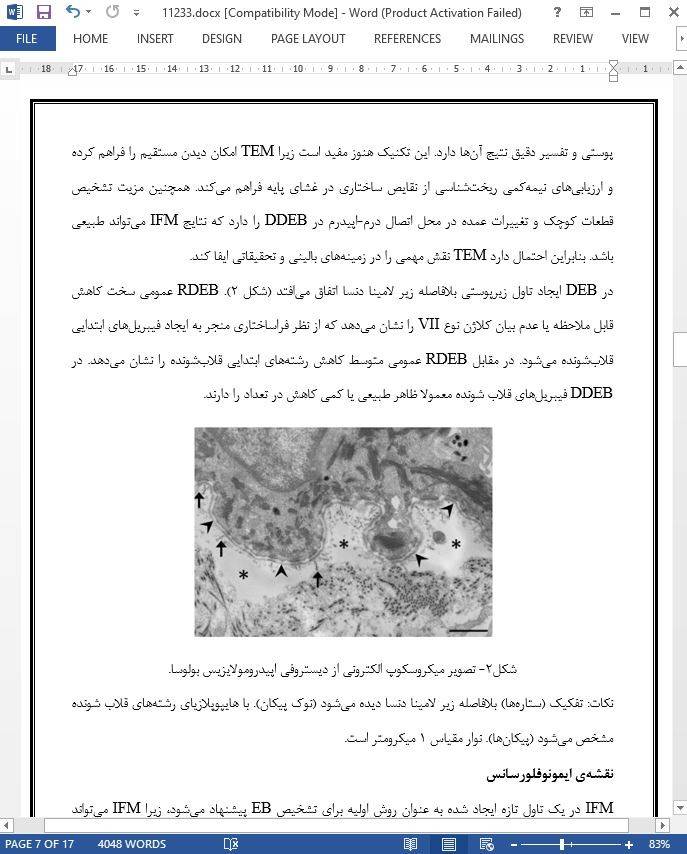
میکروسکوپ الکترونی گذاره
نقشه ی ایمونوفلورسانس
آنالیز جهش
درمان بیماران با دیستروفی اپیدرمولایزیس بولوزا
پیشرفت های درمانی
نتیجه گیری
Abstract
Introduction
Type VII collagen and the pathogenesis of dystrophic epidermolysis bullosa
Clinical features of dystrophic epidermolysis bullosa
Clinical subtypes
DDEB, generalized
RDEB, generalized severe
RDEB, generalized intermediate
Diagnosis of dystrophic epidermolysis bullosa
Transmission electron microscopy
Immunofluorescence mapping
Mutational analysis
Treatments for patients with dystrophic epidermolysis bullosa
Treatment advances
Conclusion
- اصل مقاله انگلیسی با فرمت ورد (word) با قابلیت ویرایش
- ترجمه فارسی مقاله با فرمت ورد (word) با قابلیت ویرایش، بدون آرم سایت ای ترجمه
- ترجمه فارسی مقاله با فرمت pdf، بدون آرم سایت ای ترجمه


