دانلود مقاله اجرای پردازش سیگنال دیجیتالی زمان واقعی برای in-beam PET تصویربرداری پرتودرمانی در HIMM
چکیده
برش نگاری با گسیل پوزیترون in-beam (ibPET) به تصویربرداری پرتودرمانی دستگاه پزشکی یون-سنگین (HIMM) اختصاص داده می شود، که به اندازه گیری آنلاین دقیق و همچنین توانایی پردازش سیگنال زمان واقعی الکترونیک ها نیاز دارد. ما در این مقاله اجرای پردازش سیگنال دیجیتالی زمان واقعی را در واحد اکتساب داده (DAQU) برای یک سیستم ibPET براساس بازخوانی لامپ تکثیر فوتونی (PMT) کریستال های سوسوزن اکسی اورتوسیلیکات لوتتیوم-ایتریوم (LYSO) ارائه می دهیم. ما مدار پردازش سیگنال سفارشی 10 کانالی را بر روی یک FPGA با عملکرد بالا طراحی کرده ایم، که از 3 حالت کاری (حالت پردازش سیگنال تک رویدادی، حالت خود کالیبراسیون و حالت داده خام (ADC))، محاسبه موقعیت، مکان یابی کریستالی، ذخیره سازی رویداد و غیره پشتیبانی می کند.
1. مقدمه
تعداد مراکزی که در سراسر دنیا از ذرات تراپی برای درمان سرطان استفاده می کنند، در حال افزایش است [1]، زیرا ذرات با بار سنگین رسوب انرژی انتخابی تری در در عمق و افزایش اثربخشی زیستی دارند [3، 2]. بنابراین، نظارت زمان واقعی فضای پرتو و توزیع دوز در طول درمان که می تواند ایمنی و دقت برنامه درمان را تضمین کند، به نقطه داغ در زمینه های رادیولوژی و پزشکی هسته ای تبدیل می شود. ابتدا این گروه GSI از اصطلاح روش نظارت «برش نگاری با گسیل پوزیترون in-beam (PET in-beam)» در روال بالینی استفاده می کند. یک PET In-beam برای نظارت بر دوز پرتوافکنی و ارائه دقیق موقعیت به بیمار از طریق تشخیص رویدادهای نابودی تولید شده در ناحیه تابش در طول پرتودرمانی یون و تکمیل بازسازی تصویر در مقیاس رنگی برای اعتبارسنجی درمان توسعه می یابد [7]. برخی از تیم های تحقیقاتی دیگر با تایید امکان پذیری و مقدار بالینی ibPET، هندسه های سیستم با طراحی خاص را برای برنامه های تصویربرداری بالینی با محدوده کوچک شامل آشکارساز های اختصاصی و سیستم های الکترونیکی بازخوانی توسعه داده اند [10-8].
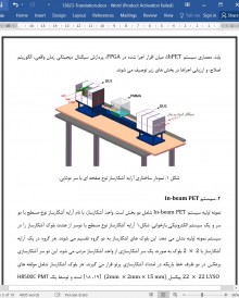
اولین نسل دستگاه پزشکی یون-سنگین (HIMM) [12، 11] برای درمان سرطان در مرکز درمان ووِی، گانسو برای کاربرد بالینی ساخته شده است. یک ibPET اختصاصی برای بهبود عملکرد دستگاه صنعتی سازی HIMM داخلی بر روی خط باریکه در HIMM بروزرسانی شده برای نظارت در شرایط غیرآزمایشگاهی و غیرتهاجمی اجرا خواهد شد. با این حال، هنوز فقدان راه حل ها برای یکپارچگی PET تجاری در یک مرکز درمان وجود دارد [14، 13]، بنابراین آشکارسازهای اختصاصی باید توسعه یابند. یک هندسه مسطح دوگانه به ویژه برای ibPET مناسب است، زیرا به پرتو این امکان را می دهد تا بدون آسیب به آشکارسازهای اسکنر PET از بدن بیمار عبور کند [6، 5]. چنین سیستم هایی در بازسازی دقیق چالش هایی دارند، که در آن تصاویر باید تحت نرخ شمارش زیاد و در یک زمان کوتاه برای امکان بازخورد ایجاد شوند. بنابراین، الکترونیک های بازخوانی آن برای داشتن قابلیت موقعیت زمان واقعی، اندازه گیری زمان و انرژی به وضوح بالا برای واحد اکتساب داده (DAQU) از ibPET ضروری است. در این مقاله، اجرای یک طرح پردازش سیگنال دیجیتالی زمان واقعی ارائه می شود، که برای واحد اکتساب داده (DAQU) از ibPET توسعه می یابد. معماری سیستم ibPET، میان افزار اجرا شده در FPGA، پردازش سیگنال دیجیتالی زمان واقعی، الگوریتم اصلاح، و ارزیابی اجراها در بخش های زیر توصیف می شوند.
5. نتیجه گیری
طراحی و اجرای پردازش و اصلاح سیگنال دیجیتالی زمان واقعی مبتنی بر FPGA در این مقاله توصیف می شود. نتایج آزمایشات عملکرد الکترونیکی نشان می دهند که طراحی تابع منطقی به درستی کار می کند. علاوه بر این، مقایسه نتایج برای پردازش زمان واقعی و تحلیل آفلاین نشان می دهند که طراحی منطقی پردازش داده زمان واقعی شرط کنترل آنلاین PET in-beam را بدست می آورد.
Abstract
The in-beam positron emission tomography (ibPET) is dedicated to radiotherapy imaging of the heavy-ion medical machine (HIMM), which requires precise online measurement, as well as real-time signal processing capability of the electronics. In this paper, we present the implementation of real-time digital signal processing in the data acquisition unit (DAQU) for an ibPET system based on the photomultiplier tube (PMT) readout of lutetium-yttrium oxyorthosilicate (LYSO) scintillator crystals. We have designed 10-channel customized signal processing circuit on a high-performance FPGA, which supports 3 working modes (single event processing mode, self-calibration mode and raw ADC data mode), position calculation, crystal locating, event sorting, and etc. Moreover, the implementation of real-time corrections is capable of dealing with photon peak correction to 511 keV and crystal ID-based time correction. The implemented real-time ibPET system can achieve 46% data compression and beyond 1,000,000 events/sec/channel signal processing capabilities. A series of initial tests is conducted, and the results indicate that this design meets the application requirement on online processing.
1. Introduction
The number of centers where particle therapy is employed for cancer treatment is increasing worldwide [1], as heavy charged particles have a more selective deposition of energy in depth and an increased biological effectiveness [2,3]. Therefore, the real-time monitoring of beam spatial and dose distribution during the treatment, which can ensure the safety and accuracy of the treatment plan, becomes the hotspot in the fields of radiology and nuclear medicine. The GSI group uses the first to use the so-called ‘in-beam positron emission tomography (In-beam PET)’ [4–6] monitoring technique in the clinical routine. An In-beam PET is developed for monitoring the irradiation dose and position accurately delivered to patient through the detection of annihilation events generated at the irradiation area during ion beam therapy, and completing the color-scale image reconstruction for treatment validation [7]. Some other research teams have developed specifically tailored system geometries for small-scoped clinical imaging applications, including dedicated detectors and readout electronic systems, verifying the feasibility and clinical value of ibPET [8–10].
The first generation heavy-ion medical machine (HIMM) [11,12] has been constructed for cancer therapy at the Wuwei, Gansu, treatment center for clinical application. To improve the performance of the domestic HIMM industrialization device, a dedicated ibPET will be mounted on the beam line in the upgraded HIMM for in vivo and non-invasive monitoring. However, there is still lack of solutions to integrate a commercial PET into a treatment center today [13,14], thus the dedicated detectors need to be developed. A dual planar geometry, which allows the beam to pass through the patient without hitting the detectors of the PET scanner, is particularly convenient for ibPET [5,6]. Such systems have challenges in accurate reconstruction, where images have to be generated under high count rate and within a short time to allow feedback. Therefore, its readout electronics is required to have the capability of real-time position, time and energy measurement with high-resolution [15–17]. In the paper, a design implementation of realtime digital signal processing is presented, which is developed for the Data Acquisition Unit (DAQU) of ibPET. The ibPET system architecture, firmware implemented in FPGA, real-time digital signal processing, correction algorithm, and performances evaluation are described in the following sections.
5. Conclusion
The design and implementation of FPGA-based real-time digital signal processing and correction is described in this paper. The results of the electronics performance tests indicates that the logic function design works properly. In addition, the comparison of results for realtime processing and off-line analysis indicates that the real-time data processing logic design achieves the online monitoring requirement of the in-beam PET.
چکیده
1. مقدمه
2. سیستم PET In-beam
3. طراحی منطق پردازشی رویداد DAQU
3.1. حالت پردازش تک رویدادی
3.2. حالت خود کالیبراسیون
3.3. حالت داده خام ADC
3.4. مقایسه و انتقال داده
4. نتایج آزمایشی و مقایسه
4.1. نتایج آزمایش عملکرد DAQU
4.1.1. رزولوشن موقعیت
4.1.2. وضوح زمان بندی
4.1.3. وضوح انرژی
4.2. مقایسه نتایج برای پردازش زمان واقعی و تحلیل آفلاین
4.2.1. مقایسه شناسایی کریستالی
4.2.2. مقایسه اصلاح اوج فوتون
5. نتیجه گیری
منابع
Abstract
1. Introduction
2. The In-beam PET system
3. Design of DAQU event processing logic
3.1. Single event processing mode
3.2. Self-calibration mode
3.3. Raw ADC data mode
3.4. Data compression and transmission
4. Testing results and comparison
4.1. Performance test results of the DAQU
4.1.1. Position resolution
4.1.2. Timing resolution
4.1.3. Energy resolution
4.2. Comparison of results for real-time processing and off-line analysis
4.2.1. Comparison of crystal identification
4.2.2. Comparison of photon peak correction
5. Conclusion
Acknowledgments
References
این محصول شامل پاورپوینت ترجمه نیز می باشد که پس از خرید قابل دانلود می باشد. پاورپوینت این مقاله حاوی 22 اسلاید و 5 فصل است. در صورت نیاز به ارائه مقاله در کنفرانس یا سمینار می توان از این فایل پاورپوینت استفاده کرد.
در این محصول، به همراه ترجمه کامل متن، یک فایل ورد ترجمه خلاصه نیز ارائه شده است. متن فارسی این مقاله در 9 صفحه (1570 کلمه) خلاصه شده و در داخل بسته قرار گرفته است.
علاوه بر ترجمه مقاله، یک فایل ورد نیز به این محصول اضافه شده است که در آن متن به صورت یک پاراگراف انگلیسی و یک پاراگراف فارسی درج شده است که باعث می شود به راحتی قادر به تشخیص ترجمه هر بخش از مقاله و مطالعه آن باشید. این فایل برای یادگیری و مطالعه همزمان متن انگلیسی و فارسی بسیار مفید می باشد.
بخش مهم دیگری از این محصول لغت نامه یا اصطلاحات تخصصی می باشد که در آن تعداد 40 عبارت و اصطلاح تخصصی استفاده شده در این مقاله در یک فایل اکسل جمع آوری شده است. در این فایل اصطلاحات انگلیسی (تک کلمه ای یا چند کلمه ای) در یک ستون و ترجمه آنها در ستون دیگر درج شده است که در صورت نیاز می توان به راحتی از این عبارات استفاده کرد.
- ترجمه فارسی مقاله با فرمت ورد (word) با قابلیت ویرایش و pdf بدون آرم سایت ای ترجمه
- پاورپوینت فارسی با فرمت pptx
- خلاصه فارسی با فرمت ورد (word)
- متن پاراگراف به پاراگراف انگلیسی و فارسی با فرمت ورد (word)
- اصطلاحات تخصصی با فرمت اکسل


