
دانلود رایگان مقاله اکسیداسیون حرارتی فیلم های غیرمتبلور و نازک فلزی Zr-Cu-Al-Ni
چکیده
مراحل ابتدایی اکسیداسیون حرارتی فیلم های نازک و غیرمتبلور فلزی Zr-CuAl-Ni با استفاده از اسپکتروسکوپی فوتوالکترونی اشعه x، میکروسکوپ الکترونی و اسپکتروسکوپی پراش اشعه x مورد بررسی قرار گرفت. فیلم های به وجود آمده دارای اکسیزن پیوندی است که به پایداری فاز غیر متبلور کمک می کند. این اکسیژن پس از حرارت دیدن تا 300 درجه در مدت زمان های کوتاه (5 دقیقه) از سطح فصل مشترک جدا می شود. حرارت دهی تا 300 درجه در مدت زمان طولانی تر موجب تجزیه افقی و عمودی و تشکیل یک لایه سطحی اکسید شامل اکسیدهای Zr و Al می شود. تشکیل اکسید سطحی در ابتدا با نفوذ معکوس Cu و Ni و (<30 دقیقه) و سپس با نفوذ بیرونی Zr (>30 دقیقه) محدود می شود. خواص اکسیداسیون تطابق بسیاری با مشاهدات قبلی شیشه های فلزی Zr-Cu-Al-Ni داشت البته اختلافاتی مشاهده شد که می توان آن ها را با شکل هندسی منحصر به فرد فیلم های نازک و غیرمتبلور فلزی توجیه کرد.
1. مقدمه
فلزات غیرمتبلور حجمی (شیشه فلزات حجمی، BMGها) در 50 ساله اخیر به طور وسیع مورد بررسی قرار گرفته اند [1]. به ویژه از بین این فلزات شیشه فلزات حجمی برپایه Zr-Cu مانند Zr-Cu-Al-Ni(ZCAN) به علت قابلیت تشکیل شیشه، قدرت مکانیکی بالاو مقاومت در برابر خوردگی بیشتر مورد توجه قرار گرفته است [1و2]. اخیرا پتانسیل فیلم های نازک و نامتبلور فلزی (AMTFs) به عنوان جایگزینی برای فیلم های نازک پلی کریستالی در بسیاری از کاربردها کشف شده است [3]. از بینAMFT ها، ZCAN برای کاربردهایی مثل الگوسازی نانو [4و5]، پوشش دهی مکانیکی [6]، نانو ورقه ها [7و8] و دستگاه های فلز- جداکننده- فلز [9و10] مورد بررسی کامل قرار گرفته است.
شناخت اکسیداسیون حرارتی این فلزات برای کاربردهای ذکر شده و سایر کاربردهای بالقوه اساسی است. به عنوان مثال ته نشینی لایه های دی الکتریک برای دستگاه های فلز-جداکننده-فلز و بسیاری از کاربردهای مشابه به دماهایی بالاتر از 300 درجه نیاز دارد [9]. اکسیداسیون AMTF ها می تواند هم منجر به تخریب و هم منجر به بهبود خواص مواد گردد. به عنوان مثال در عین حال که اکسیداسیون برای کاربردهای میکروالکترونیک مناسب نیست چون به مقاومت کم و تعریف شده ای نیاز است، اما در وسایل بیودرمانی به عنوان لایه ای غیرفعال مناسب است و این کاربرد هم برای فیلم نازک و نامنظم [12] و هم برای فلزات حجمی [11] مناسب می باشد. با وجود اینکه مطالعات چشمگیری برای درک بهتر اکسیداسیون ZCAN به عنوان یک BMG انجام شده اما این مطالعات به شدت به زمان های طولانی مدت حرارت دهی محدود شده که منجر به نفوذ چشمگیر همه گونه های فلزی می گردد. درک اکسیداسیون ابتدایی (و به دنبال آن تغییر ترکیب) AMTF هایی مثل ZCAN [13] چندان مورد توجه قرار نگرفته و انتظار می رود بسیار مورد علاقه مجامع علمی باشد.
علاوه بر اکسیداسیون حرارتی نقش مقادیر اندک اکسیژن که در حین سنتز مواد ترکیب می شود برای بسیاری از فلزات نامنظم شامل ZCAN در هردو شکل فیلم نازک[14]و حجمی [14] مورد مطالعه قرار گرفته و نشان داده شده که ترکیب اکسیژن تاثیرات بسیاری بر خواص فیزیکی و ساختاری دارد [16]. در نتیجه پایداری حرارتی اکسیژن در این فیلم ها برای بسیاری از کاربردها جزء ملزومات فرایند است.
در این مطالعه اکسیداسیون حرارتی فیلم های نازک و غیرمتبلور فلزی ZCAN با استفاده از اسپکتروسکوپی فوتوالکترونی اشعه (XPS)x، میکروسکوپ الکترونی (TEM)و اسپکتروسکوپی پراش اشعه x (EDS)مورد بررسی قرار گرفت. نتایج ما چشم اندازی از مراحل ابتدایی اکسیداسیون فیلم های نازک ACAN در طول دوره حرارت دهی تا 300 درجه درهوا را به دست می دهد.
2. جزییات آزمایشی
فیلم های نازک ZCAN (با ضخامت 50 نانومتر) روی قطعه های اکسید شده Si (SiO2 با ضخامت 140 نانومتر) و پنجره های SiN(برای آنالیز TEM) قرارداده شد. برای نشاندن فیلمهای ZCAN از کندوپاش مگنترون با استفاده از Zr40Cu35Al15Ni10 به ضخامت 3 اینچ، فشار 3 میلی تور و دبی جریان آرگون برابر 20 sccm و فاصله هدف گیری 4 اینچ استفاده شد. نمونه های بازنشانی شده با استفاده از کوره تونلی در حضور هوا اکسید شدند. نمونه ها در یک کوره تونلی که در دمای محیط نگه داشته شده بود قرار داده شد. بعد از اکسیداسیون در شرلیط دلخواه (5، 15، 30، 60 و 120 دقیقه در دمای 300 درجه و 60 دقیقه در دمای 400 درجه) نمونه ها در دمای محیط سرد شد.
برای آنالیز TEM از یک میکروسکوپ الکترونیFEI 80-200 kV Titan که در 200kV کار می کند و اسپکتروسکوپی پراش اشعه ایکس (EDS)ChemiSTEM استفاده شد. نمونه ها با مستقیم قراردادن فیلم ها برروی شبکه های TEM با پنجره های نازک 200 نانومتری SiN و یا به صورت فیلم های نازکی که با استفاده از پرتو متمرکز یونی (FIB) در میکروسکوپ الکترونی FEI 3D ایجاد می شود، آماده گردید. خطوط اسکن EDS، باطول گام بین 1 و 2 نانومتر جمع آوری شد و برای کاهش خطا، داده ها حول 5 داده میانگین گرفته شد.
اندازه گیری های XPS با سیستم پویش PHI و با استفاده از پرتوتابی تک رنگ AlKα با اندازه 100 میکرومتر انجام شد. داده ها با زاویه انحراف 45 درجه و انرژی عبور 140eV برای پروفایل کندوپاش به دست آمد. مقیاس انرژی اسپکترومتر بر اساس Au 4f7/2 در eV84 و Cu 2p3/2 در eV7/932 تنظیم شده است. پروفایل عمق کندوپاش با کندوپاش متناوب نمونه و سپس به دست آوردن داده ها با دقت بالا در هر چرخه کندوپاش حاصل شد. زمان های کندوپاش XPS برای تخمین عمق ها با ارتباط دادن عکس های TEM فیلم هایی که به مدت 60 دقیقه تا 300 درجه حرارت دهی شده بود، تنظیم گردید. در این مورد نرخ کندوپاش به صورت جداگانه برای فیلم فلزی و اکسید سطحی تعیین شد. محاسبات کمی سازی که با استفاده از PHI Multipak که فاکتورهای حساسیت تابع انتقال آنالیزور و مقادیر گزارش شده را به هم پیوند میدهد بایستی شبه کمی در نظر گرفته شود. بررسی های عمق پروفایل حالت شیمیایی در هر چرخه کندوپاش با تطبیق طیف سه بعدی Zr در اجزاء فلزی (فلز Zr) و اکسیدی (اکسید Zr) انجام شد. کمی سازی پروفایل های عمق کندوپاش در اثرات کندوپاش حرفه ای به حساب نیامد.
3. بحث و نتیجه گیری
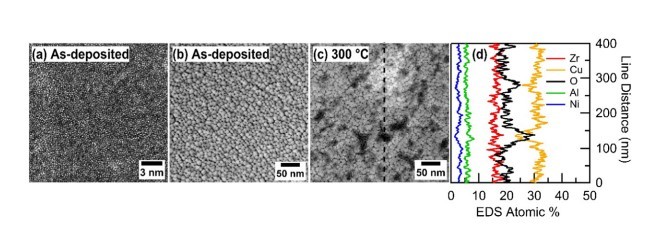
تصویر آنالیز TEM فیلم های ZCAN قبل از 60 دقیقه حرارت دهی در 300 درجه در شکل 1 نشان داده شده است. در شکل 1a یک تصویر زمینه سفید با رزولوشن بالا (HRTEM) از فیلم لایه نشانی شده نشان داده شده است. همان طور که در مطالعات پیشین هم تایید شده بود تصاویر نشان می دهد که فیلم ها ساختاری غیر بلوری و نامنظم دارند [9]. در شکل 1b تصویر HAADT فیلم نشان داده شده است. در این تصویر دیده می شود که مناطق با دانسیته الکترونی بالا ( مناطق روشن) با درجه بزرگی 10 نانومتر توسط مناطق با دانسیته الکترونی پایین (مناطق پررنگ) محاصره شده است. ناهمگونی های کوچکی که در تصویر HAADF دیده می شود می تواند به علت وجود تخلخل در ساختار فیلم و یا جدایی عناصر با دانسیته الکترونی پایین (مثل اکسیژن) از مرزهای فلزی باشد. شکل 1c تصویر HAADF فیلم ZCAN را پس از 60 دقیقه حرارت دهی در 300 درجه در هوا نشان می دهد. این شرایط حرارت دهی باعث کاهش خوشه ای شدن نمونه ها و توسعه ناهمگونی ها می شود. ای ناهمگونی ها به صورت نقاط روشن و تیره با اندازه 10 تا 100 نانومتر به صورت پخش شده روی فیلم ZCAN دیده می شود. برای دستیابی به چشم اندازی درمورد اختلاف ترکیبی ممکن بین مناطق تیره و روشن داده های EDS خط چین روی شکل c را در شکل 1d نشان داده ایم. خط اسکن نشان میدهد که مناطق تیره در فیلم نشان دهنده مناطق غنی از اکسیژن منطبق بر پیک غلظت O در فاصله 40، 135، 280 و 390 نانومتر از خط است. همچنین در این نواحی کاهش چشمگیر در محتوی Cu و کاهش تقریبی در محتوی Ni دیده می شود که نشان می دهد این نواحی از اکسیدهای Zr و Al تشکیل شده است. خط اسکن EDS همچنین ناحیه روشنی را در فاصله 300 نانومتری خط نشان می دهد. اختلاف چشمگیری در ترکیب این رژیم دیده نمی شود بنابراین روشن بودن این ناحیه به علت افزایش ضخامت فیلم است. بنابراین در طول گرمادهی در 300 درجه ضخامت تغییر میکند.
The initial stages of thermal oxidation for Zr–Cu–Al–Ni amorphous metal thin films were investigated using X-ray photoelectron spectroscopy, transmission electron microscopy and energy dispersive X-ray spectroscopy. The asdeposited films had oxygen incorporated during sputter deposition, which helped to stabilize the amorphous phase. After annealing in air at 300 °C for short times (5 min) this oxygen was found to segregate to the surface or buried interface. Annealing at 300 °C for longer times leads to significant composition variation in both vertical and lateral directions, and formation of a surface oxide layer that consists primarily of Zr and Al oxides. Surface oxide formation was initially limited by back-diffusion of Cu and Ni (b30 min), and then by outward diffusion of Zr (N30 min). The oxidation properties are largely consistent with previous observations of Zr–Cu–Al–Ni metallic glasses, however some discrepancies were observed which could be explained by the unique sample geometry of the amorphous metal thin films.
1. Introduction
Bulk amorphous metals (i.e., bulk metallic glasses, BMGs) have been extensively investigated over the last 50 years [1]. In particular, Zr–Cubased BMGs such as Zr–Cu–Al–Ni (ZCAN) have attracted significant interest due to their good glass forming ability, high mechanical strength and corrosion resistance [1,2]. More recently, amorphous metal thin films (AMTFs) have been shown to have potential as a replacement for polycrystalline thin films for a wide range of applications [3]. ZCAN is among the most thoroughly investigated AMTFs for applications such as nanoscale patterning [4,5], mechanical coatings [6], nanolaminates [7,8] and metal-insulator-metal devices [9,10].
Understanding the thermal oxidation of these materials is critical for the aforementioned and other potential applications. For example, the deposition of dielectric layers for metal-insulator-metal (MIM) devices often requires temperatures N 300 °C [9], and many other applications require similar thermal processes. The oxidation of AMTFs may either improve or degrade the materials properties. For example, while oxidation may be unfavorable for microelectronic applications where low resistivity and a well-defined interface are required, it may be desirable as a passivation layer for biomedical devices, which is an emerging application for both bulk [11] and thin film [12] amorphous metals. Although significant research has been conducted to better understand the oxidation of ZCAN as a BMG, these studies are largely limited to long annealing times at high temperatures which allows significant diffusion of all metal species. Understanding the initial oxidation (and associated composition variation) of AMTFs such as ZCAN [13] has received considerably less attention and is expected to be of interest to the scientific community at large.
In addition to thermal oxidation, the role of small quantities of oxygen incorporated during the material synthesis has been studied for many amorphous metals, including ZCAN in both thin film [14] and bulk [2,15] forms, and incorporation of oxygen has been shown to have significant effects on both structural and physical material properties [16]. Thus, the thermal stability of oxygen in these films has implications related to the processing requirements for many applications.
In this study the thermal oxidation of amorphous oxygen-containing ZCAN thin films was investigated using X-ray photoelectron spectroscopy (XPS), transmission electron microscopy (TEM) and energydispersive X-ray spectroscopy (EDS). Our results provide insight into the initial stages of thermal oxidation for ZCAN thin films during a 300 °C anneal in air.
2. Experimental details
ZCAN thin films (≈50 nm thick) were deposited onto thermally oxidized Si wafers (SiO2 thickness ≈ 140 nm) and SiN windows (for topdown TEM analysis). DC magnetron sputtering was used to deposit the ZCAN films using a 3-in. Zr40Cu35Al15Ni10 target (Kamis Incorporated), a power of 60 W, a pressure of 3 mTorr, an Ar flow rate of 20 sccm, and a target to substrate distance of 4 in. The as-deposited samples were thermally oxidized in air using a tube furnace. The samples were inserted into a tube furnace held at the target temperature. After oxidation at the desired conditions (5, 15, 30, 60 and 120 min at 300 °C and 60 min at 400 °C) the samples were removed and allowed to cool to room temperature.
An FEI 80–200 kV Titan Scanning/Transmission Electron Microscope (S/TEM) operating at 200 kV with ChemiSTEM energy dispersive X-ray spectroscopy (EDS) was used for TEM analysis. Samples were prepared by depositing the films directly onto TEM grids with ≈20 nm thick SiN windows, or fabricated as thin film lift outs using a focused ion beam (FIB) in an FEI 3D DualBeam Scanning Electron Microscope. EDS line scans were collected with a step size between 1 and 2 nm and the data was averaged over 5 points to reduce noise.
XPS measurements were performed with a PHI Quantera Scanning ESCA system using monochromatic Al Kα radiation with a 100 μm spot size. The data were acquired with a 45° emission angle and an electron analyzer pass energy of 140 eV for the sputter depth profile data. The energy scale of the spectrometer is calibrated to Au 4f7/2 at 84.0 eV and Cu 2p3/2 at 932.7 eV. The sputter depth profiles were acquired by alternately sputtering the sample and then acquiring high resolution data at each sputter cycle. A monoatomic 2 kV Ar+ ion beam rastered over a 2 × 2 mm2 area was used for ion milling. XPS sputter times were adjusted to approximate depths by correlating to TEM cross-sectional images of films annealed at 300 °C for 60 min, where approximate sputter rates were determined separately for the metal film and the surface oxide. Quantification calculations were made using PHI MultiPak which incorporates established sensitivity factors corrected for the transmission function of the analyzer and the reported values should be regarded as semi-quantitative. Chemical state resolved depth profile analysis was performed by fitting the Zr 3d spectra into both metallic (Zrmetal) and oxide (Zr-oxide) components at each sputter cycle. The quantification of the sputter depth profiles does not take into account effects of preferential sputtering.
3. Results and discussion
Plan-view TEM analysis of ZCAN films as-deposited and after a 300 °C, 60 min anneal is shown in Fig. 1. A high-resolution bright field (HRTEM) image of the as-deposited film is shown in Fig. 1a. The salt-and-pepper pattern and the lack of long range order suggest that the films are amorphous, consistent with previous studies [9]. A high-angle annular dark field (HAADF) image of the as-deposited film is shown in Fig. 1b, and reveals high electron density (light contrast) regions on the order of 10 nm surrounded by low electron density (dark contrast) regions. The shortrange inhomogeneities observed in this low resolution HAADF image may be due to slight porosity in the film structure or segregation of low electron density elements (e.g., oxygen) to the boundaries of metal clusters. EDS analysis of this as-deposited film (data not shown) revealed a uniform 2-dimensional composition of all metal species within the spatial resolution of the technique. Fig. 1c shows a HAADF image of the ZCAN film after annealing to 300 °C for 60 min in air. These annealing conditions resulted in reduction in the clustering seen in the as-deposited sample and the development of significant long-range inhomogeneities. These inhomogeneities are seen as light and dark regions ranging from ≈10 to 100 nm that are dispersed throughout the ZCAN film. To gain insight into possible compositional variation between the dark and light regions, in Fig. 1d we show image contrast EDS data along the dashed line shown in Fig. 1c. The line scan suggests that the dark regions in the 300 °C annealed film represent oxygen-rich regions as indicated by peaks in O concentration near line distances of 40, 135, 280 and 390 nm. These regions also have a significant reduction in Cu content and a slight reduction in Ni content, suggesting these regions are composed primarily of Zr- and Al-oxides. The EDS line scan also includes a region of light contrast centered near the line distance of 300 nm. No significant change in composition is observed through this regime suggesting the lighter contrast is likely due to an increase in film thickness. Thus, thickness variation is introduced during the 300 °C anneal. It should be noted that the Cu concentration measured by EDS for this analysis was artificially increased via scattering effects from Cu present in the TEM specimen holder, however this does not affect the trends observed in the line scan.
چکیده
1. مقدمه
2. جزییات آزمایشی
3. بحث و نتیجه گیری
4. نتیجه گیری
منابع
Abstract
1. Introduction
2. Experimental details
3. Results and discussion
4. Conclusions
References