
دانلود رایگان مقاله آستاگزانتین به عنوان یک عامل محافظ اعصاب
چکیده
بیماری های عصبی، که شامل آسیب های حاد و مزمن نورودژنراتیو می شود، عامل مرگ و ناتوانی انسان می باشند. با این وجود، پاتوفیزیولوژی این بیماری ها هنوز به طور کامل روشن نشده است، و هنوز درمان موثری وجود ندارد. آستاگزانتین؛ عضوی از گروه xanthophyll، نوعی کارتنوئید قرمز-نارنجی با فعالیت های مختلف بر غشاء سلولی و فعالیت های بیولوژیکی متنوع است. مهم تر از همه، شواهدی وجود دارد که نشان می دهد آستاگزانتین دارای اثرات محافظ اعصاب در مدل های آزمایشگاهی آسیب حاد، اختلالات نورودژنراتیو مزمن، و بیماری های عصبی است. اثرات مفید آستاگزانتین در ارتباط با خاصیت ضد اکسیداتیو، ضد التهابی و ضد آپاپتوزیس است. در این مقاله مروری، ما بر روی ویژگی های محافظ اعصاب آستاگزانتین تمرکز خواهیم کرد و مکانیسم های مربوطه به آن را در بیماری های عصبی بررسی خواهیم کرد.
1. مقدمه
بیماری های عصبی، به عنوان مثال آسیب های حاد (مانند، سکته و جراحت های ضربه ای مغز) و تخریب مزمن اعصاب (مانند، بیماری آلزایمر، پارکینسون و هوتینگتون)، عامل اصلی مرگ و ناتوانی انسان هستند. استرس اکسیداتیو، التهاب، و آپاپتوزیس برخی از مکانیسم های دخیل در پاتوژنز این بیماری ها هستند. به عنوان مثال، توده های آمیلوئید بتا پپتید به شدت غیر محلول و توپی های فیبریل عصبی عامل اصلی ایجاد استرس اکسیداتیو و التهاب در مغز افراد مبتلا به آلزایمر (AD) هستند که به طور چشمگیری به مرگ نورون ها در این بیماری منجر می شوند. بعلاوه، شواهدی وجود دارند که نشان می دهد نقص میتوکندریایی، استرس های اکسیداتیو و نیتروزاتیو، تجمع و گردآمدگی پروتئین های بد ریخت و نامعمول (مانند α-synuclein) و عملکرد نادرست سیستم ubiquitin-proteasome، مکانیسم مولکولی اصلی دخیل در پاتوژنز بیماری پارکینسون (PD) تک گیر و فامیلی است. بعلاوه، تکه کاملا چند ریخت CAG tri-nucleotide در اکسون-1 ژن huntingtin تکه نامعمول و طویل poly-glutamine را کد می کند که با آسیب های مغزی ناشی از بیماری هانتینگتون (HD) مرتبط است. گسترش پلی-گلوتامین منجر به بدتر شدن هانتینگتین و تجمع آن در هسته می شود. این امر منجر به میانکنش های نامساعد با سایر پروتئین ها می شود که به تجمع داخل هسته ای موتان های هانتیگتین و تشکیل توده های نوروپیل می انجامد که نهایتا بعث مرگ سلول عصبی می شود. بنابراین، عوامل دارویی چند هدفه ممکن است برای درمان این بیماری های تحلیل برنده موثر باشد.
آستاگزانتین، یک عضو بی همتای xanthophylls، نوعی ماده مغذی گیاهی است که می تواند توسط ریز جلبک Haematococcus pluvialis سنتز شود. برخلاف سایر اعضای خانواده xanthophylls، آستاگزانتین دارای دو گروه هیدروکسیل است. آستاگزانتین در کنار دو لایه لیپید قرار می گیرد و به اندازه کافی بلند است تا دو گروه هیدروکسیل در کنار فاز مایع قرار گیرد و هنگامی که الکترون ها از این گروه های هیدروکسیل توسط رادیکال های آزاد جدا شدند، رزونانس مولکولی پایدار باشد. در نتیجه، این ویژگی ها اجازه می دهد تا آستاگزانتین تا کارهای زیادی را در بدن انجام دهد. به عنوان مثال، آستاگزانتین می تواند به طور چشمگیری خطر ابتلا به بیماری قلبی-عروقی را کاهش دهد. یک جیره حاوی آستاگزانتین (75 یا 200 mg/kg در روز) به مدت 8 هفته باعث بهبود اتصاع عروقی وابسته به آندوتلیوم در عروق مقاوم شد، فشار خون کاهش یافت، و تغییر شکل قلبی در رتهای مبتلا به فشار خون بالا برطرف گردید. علاوه بر این، آستاگزانتین(100 و 500 میلی گرم در 100 گرم) به مدت 60 روز از اکسیداسیون پروتئین سرم در خرگوش های هایپر-کلسترلمی جلوگیری کرد. همچنین مطالعات نشان داده اند که آستاگزانتین می تواند به راحتی از سد خونی-مغزی عبور کند تا مغز را در برابر جراحت های نورودژنراتیو حاد و مزمن محافظت نماید. ویژگی های محافظت از اعصاب این مولکول شامل، ضد-اکسیداسیون، ضد التهاب، و ضد-آپاپتوزیس است. بنابراین این مقاله مروری بر روی اثرات مفید آستاگزانتین متمرکز خواهد شد و مکانیسم های دخیل مشاهده شده در مدل های آزمایشگاهی بیماری های عصبی را بررسی خواهد کرد.
2. آستاگزانتین: منبع، بیوشیمی، فراهم زیستی، و سلامتی
Xanthophyll گروهی از رنگ دانه های کارتنوئیدی حاوی اکسیژن است که در گیاهان سنتز شده و از لیکوپن مشتق می گردد. آستاگزانتین یک رنگدانه قرمز است که به این خانواده تعلق دارد. این ترکیب به طور طبیعی در طیف وسیعی از ارگانیسم های زنده شامل، میکروجلبک ها، گیاهان پیچیده و غذاهای دریایی وجود دارد. شکل تجاری آستاگزانتین اغلب از جلبک Haematococcus pluvialis و مخمر Phaffia rhodozyma سنتز می شود. به عنوان عضوی از گروه xanthophyll، آستاگزانتین بسیار مرتبط با سایر کرتنوئید ها مانند بتا-کاروتن، لوتئین و گزانتین است. به طور مشابهی، آنها دارای خواص فیزیولوژیک و متابولیک مشترکی دارند. برخلاف بتا-کاروتن، آستاگزانتین دارای خاصیت پیش ساز ویتامین A در بدن نیست.
جرم مولی آستاگزانتین 596.84 g/mol است و فرمول مولکی آن C40H52O4 می باشد. آن نوعی مولکول متقارن حاوی دو حلقه انتهایی متصل به یک حلقه کوتاه پلی ان است. گروه هیدروکسیل در انتهای مولکول آن را قادر می سازد تا اسیدهای چرب را استریفیه کند تا مونو و دی استرها به وجود آیند. آستاگزانتین طبیعی عمدتا به شکل استریفیه وجود دارد، در حالی که شکل مصنوعی آن به فرم آزاد تولید می شود. همچنین آستاگزانتین دارای باندهای دوگانه کونژوگه است که به این مولکول از طریق اهداء الکترون خاصیت آنتی-اکسیدانی قوی می دهد و با رادیکال های آزاد وارد واکنش می شود تا زنجیره واکنش داخلی سلولی را متوقف سازد.
آستاگزانتین دارای هر دو خصوصیت آبدوستی و آبگریزی است، بنابراین آن قابل حل در چربی می باشد و می تواند، بوسیله مولکول های چربی به بافت ها و ارگان هایی که به آن بیشترین نیاز را دارند، مانند مغز، شبکیه و عضلات اسکلتی به طور مستقیم حمل شود. آستاگزانتین، در ابتدا از طریق انتشار ساده، به سلول های روده جذب می شود، و سپس در حضور لیپیدها تحت انتشار تسهیل یافته قرار می گیرند. اشکال غیر استریفیه به شیلومیکرون ها ملحق شده و از طریق سیستم لنفاوی به کبد منتقل می شود. کبد، این مولکول ها را به طور بیوشیمیایی به ویتامین A تبدیل نمی کند. در عوض، آن به لیپوپروتئین ها ملحق شده و از طریق گردش خون به ارگان ها و بافت ها منتقل می شود.
آستاگزانتین برای مصرف بهمراه غذا ایمن است، و تاکنون گزارشی از عوارض ناخواسته در ارتباط با آن وجود ندارد. یک مطالعه کارآزمایی بالینی دریافت که روزانه 6 میلی گرم از آستاگزانتین می تواند به طور ایمن توسط بزگسالان سالم، مصرف شود. بعلاوه، چندین مطالعه کارآزمایی بالینی نشان داده است که عصاره غنی از آستاگزانتین Haematococcus pluvialis همچنین ایمن و بی خطر است. Hoffman-La Roche ایمنی آستاگزانتین در تست های حاد، جهش زایی، ناقص الخلقه زایی، سمیت رویان و سمیت دستگاه تولید مثل اثبات کردند. همچنین، سازمان غذا و داروی آمریکا استفاده از آستاگزانتین را به عنوان مکمل غذایی در سال 1999 تایید کرد.
3. ویژگی های محافظت از عصب آستاگزانتین در بیماری های عصبی
مطالعات بسیاری در ارتباط با اثرات مفید آستاگزانتین وجود دارد. محافظت از عصب ناشی از آستاگزانتین در مدل های آزمایشگاهی اختلالات عصبی شامل مکانیسم های ضد-اکسیدانی، ضد-التهابی، و ضد0 آپاپتوزیس است. این قسمت از مقاله به این مکانیسم های مولکولی خواهد پرداخت و از آنها به عنوان درمان بالقوه بیماری های عصبی یاد خواهد کرد.
3.1 تاثیر آنتی-اکسیدانتی
استرس اکسیداتیو یک میانجی اصلی در پاتولوژی بیماری های عصبی است. اختلال در تعادل وضعیت واکنش های پیش-اکسیدانتی/آنتی-اکسیدانتی در سلول ها منجر به استرس اکسیداتیو می شود که باعث تولید ریشه های فعال اکسیژن (ROS) و رادیکال های آزاد می گردد. هنگامی که این موارد در مقدار زیاد تولید می شوند، ROS مانند آنیون سوپراکسید (O2-) و محصول تجزیه آن، هیدروژن پراکسید (H2O2) برای عملکردهای متابولیک مضر هستند. رادیکال سوپراکسید می تواند آهن و گوگرد را اکسید کند و مانع آزاد سازی یون آهن شود. بنابراین یون آهن با هیدروژن پراکسید واکنش می دهد تا رادیکال بسیار قوی هیدروکسیل به دست آید. این مواد با سوبستراهای ارگانیک مانند DNA، پروتئین ها و لیپیدها واکنش های دیگری را ایجاد می کنند که منجر به اختلال در عملکرد و مرگ سلول می شود. لازم به ذکر است که آستاگزانتین می تواند به عنوان یک سپر علیه آسیب اکسیداتیو از طریق مکانیسم های مختلف مانند سرکوب اکسیژن تک الکترونی، حذف رادیکال های آزاد، ممانعت از پراکسیداسون چربی و تنظیم بیان ژن های مرتبط با استرس اکسیداتیو عمل کند. به عنوان مثال، آستاگزانتین اثرات مفیدی علیه آسیب کلیوی القاء شده توسط هیدروکلرید جیوه از طریق جلوگیری از اکسیداسشون چربی و پروتئین اعمال می کند. یک مدل درون تنی حیوانی، نشان داد که تجویز آستاگزانتین مانع آسیب ناشی از N-Methyl-D-aspartate می شود که با کاهش پراکسیداسیون چربی و اکسیداسون DNA در ارتباط است. تیمار با آستاگزانتین استرس اکسیداتیو ناشی از cyclophosphamide و آسیب DNA متعاقب را در هپاتوسیت های موش رت کاهش می دهد. اثرات محافظتی این مولکول با فعالسازی فاکتور شماره 2 وابسته به هسته اریتروسیت (Nrf2) در ارتباط است که به واقع بیان ژن های وابسته به Nrf2 مانند heme oxygenase-1 و NAD(P)H، quinine oxidoreductase-1 را تسهیل می کند. در رنگدانه اپیتلیا شبکیه انسان (RPE) رده سلولی ARPE-19، آستاگزانتین مانع از تولید داخل سلولی ROS می شود و از کاهش زنده مانی رنگدانه های سلولی در اثر H2O2 جلوگیری می کند. همچنین آستاگزانتین جابجایی هسته ای Nrf2 را افزایش می دهد و بیان ژن های فاز 2 آنزیم های آنتی اکسیدانتی را از طریق فعالسازی مسیر فسفواینوزیتول 3-کیناز (PI3K) افزایش می دهد که نهایتا به محافظت از سلول ARPE-19 در برابر استرس اکسیداتیو ناشی از H2O منجر می گردد.
Abstract
Neurological diseases, which consist of acute injuries and chronic neurodegeneration, are the leading causes of human death and disability. However, the pathophysiology of these diseases have not been fully elucidated, and effective treatments are still lacking. Astaxanthin, a member of the xanthophyll group, is a red-orange carotenoid with unique cell membrane actions and diverse biological activities. More importantly, there is evidence demonstrating that astaxanthin confers neuroprotective effects in experimental models of acute injuries, chronic neurodegenerative disorders, and neurological diseases. The beneficial effects of astaxanthin are linked to its oxidative, anti-inflammatory, and anti-apoptotic characteristics. In this review, we will focus on the neuroprotective properties of astaxanthin and explore the underlying mechanisms in the setting of neurological diseases.
1. Introduction
Neurological diseases, exemplified by acute injuries (e.g., stroke and traumatic brain injury) and chronic neurodegeneration (e.g., Alzheimer’s disease, Parkinson’s disease, and Huntington’s disease), are common causes of human death and disability [1,2]. Oxidative stress, inflammation, and apoptosis are some of the mechanisms involved in the pathogenesis of these diseases [3,4]. For example, highly insoluble amyloid beta peptide deposits and neurofibrillary tangles provide obvious stimuli for oxidative stress and inflammation in a brain with Alzheimer’s disease (AD), which significantly contributes to neuronal death in this disease [5–7]. In addition, there is evidence demonstrating that mitochondrial deficits, oxidative and nitrosative stress, accumulation and aggregation of aberrant or misfolded proteins (i.e., α-synuclein), and dysfunction of ubiquitin-proteasome system represents the principal molecular events that commonly underlie the pathogenesis of familial and sporadic forms of Parkinson’s disease (PD) [8,9]. Additionally, highly polymorphic CAG tri-nucleotide repeat expansions in exon-1 of the huntingtin gene encodes an abnormally long poly-glutamine repeat, which is associated with Huntington’s disease (HD)-related brain pathology [10]. Poly-glutamine expansion causes huntingtin to aggregate and accumulate in the nucleus. This leads to abnormal interactions with other proteins, which results in intra-nuclear accumulation of mutant huntingtin and the formation of neuropil aggregates that may ultimately lead to neuronal cell death [11]. Therefore, multi-targeted pharmacological agents may be effective for the treatment of these devastating diseases.
Astaxanthin, a unique member of the xanthophylls, is a deep red-colored phytonutrient that can be synthesized by a microalgae called Haematococcus pluvialis [12]. Distinct from other members of the xanthophylls, astaxanthin has two hydroxyl groups [13]. Astaxanthin spans the bi-lipid layer and is long enough that the two hydroxyl groups jut into the fluid phase near the membrane, and that when electrons are extracted from these hyroxyl groups by free radicals, the molecule is resonance stabilized. As a consequence, these properties allow astaxanthin to do a lot in the body. For instance, astaxanthin can dramatically decrease the risk of cardiovascular disease [14]. A diet supplemented with astaxanthin (75 or 200 mg/kg body weight per day) for 8 weeks has been shown to improve endothelium-dependent vasodilatation in resistance vessels, reduce systolic blood pressure, and improve cardiovascular remodeling in spontaneously hypertensive rats [15]. In addition, astaxanthin (100 and 500 mg/100 g) for 60 days protects against serum protein oxidation in hyper-cholesterolemic rabbits [16]. Studies have also demonstrated that astaxanthin can easily cross the BBB to protect the brain from acute injury and chronic neurodegeneration [17,18]. The neuroprotective properties of this molecule involves anti-oxidation, anti-inflammation, and anti-apoptotis [19–21]. Thus, this review article will focus on the beneficial effects of astaxanthin and explore the underlying mechanisms observed in experimental models of neurological diseases. We also propose that further studies involving astaxanthin are needed, in order to evaluate its potential application in the treatment of neurological disorders.
2. Astaxanthin: Source, Biochemistry, Bioavailability, and Safety
Xanthophyll is a class of oxygen-containing carotenoid pigments whose biosynthesis in plants derives from the lycopene. Astaxanthin is a reddish pigment which belongs to the xanthophyll family [22].This compound naturally exists in a wide variety of living organisms which includes microalgae, complex plants, and seafood [23]. The commercial form of astaxanthin is mainly synthesized from the algae Haematococcus pluvialis and the yeast Phaffia rhodozyma. As a member of the xanthophyll group, astaxanthin is closely related to other carotenoids such as β-carotene, lutein, and zeaxanthin. As a member of the xanthophyll group, astaxanthin is closely related to other carotenoids such as β-carotene, lutein, and zeaxanthin [24]. Similarly, they share many of the physiological and metabolic functions attributed to carotenoids [25]. Unlike β-carotene, astaxanthin does not have pro-vitamin A activity in the human body [26].
The molar mass of astaxanthin is 596.84 g/mol and the molecular formula is C40H52O4. It is a symmetric molecule consisting of two terminal rings joined by a short polyene ring [22]. The hydroxyl group at the end of the molecule enables it to esterify fatty acids to form mono-esters or di-esters [13]. Natural astaxanthin mainly exists in an esterified form, while the synthetic form is produced in a free form [27]. Astaxanthin also contains conjugated double bonds, giving this molecule strong anti-oxidant properties by donating electrons and reacting with free radicals to terminate free radical chain reactions within cells [25,28].
Astaxanthin has both lipophilic and hydrophilic properties, since it is fat-soluble and can be carried by fat molecules directly to tissues and organs that need it the most, like the brain, retina, and skeletal muscle [22]. Astaxanthin is first absorbed into enterocytes through passive diffusion and undergoes facilitated diffusion in the presence of lipids [29]. The unesterified forms are incorporated into chylomicrons and are transported into the liver via the lymphatic system [30]. The liver does not biochemically convert these molecules into vitamin A [31]. Instead, it is incorporated into lipoproteins that are transported into organs and tissues via the circulation [32].
Astaxanthin is safe to consume with food and contains no reports of side effects [33,34]. One randomized clinical trial found that 6 mg/day of astaxanthin can be safely consumed by healthy adults [35]. In addition, numerous human clinical trials have shown that the astaxanthin rich extract, Haematococcus pluvialis, is safe as well [36,37]. Hoffman-La Roche confirmed the safety of astaxanthin with acute, mutagenicity, teratogenicity, embryotoxicity, and reproductive toxicity tests [38]. In addition, the United States Food and Drug Administration approved the use of astaxanthin as a dietary supplement in 1999 [25].
3. Neuroprotective Properties of Astaxanthin in Neurological Diseases
There have been numerous studies concerning the beneficial effects of astaxanthin. Astaxanthin-mediated neuroprotection in experimental models of neurological disorders involves anti-oxidantion, anti-inflammation, and anti-apoptotic mechanisms [17,39]. The following sections will delve into these molecular mechanisms and their potential as treatments for neurological diseases.
3.1. Anti-Oxidant Effects
Oxidative stress is a key mediator in the pathology of neurological diseases [40–42]. Disturbance of the equilibrium status of pro-oxidant/anti-oxidant reactions in cells can lead to oxidative stress, which causes generation of reactive oxygen species (ROS) and free radicals [43]. When produced in excessive amounts, ROS like the superoxide anion radical (O2 −) and its dismutation product, hydrogen peroxide (H2O2), are detrimental to metabolic functions [44,45]. The O2 − radical can oxidize the [4Fe-4S] clusters of dehydratases, such as aconitase, causing inactivation and release of Fe2+ [46,47]. Thereafter, Fe2+ reacts with H2O2 to yield the potent oxidizing free radical species hydroxyl radical (OH). These substances further react with key organic substrates, such as DNA, proteins, and lipids, to disturb cell function and cause cell death [48]. It is worth mentioning that astaxanthin can act as a safeguard against oxidative damage through various mechanisms, by quenching of singlet oxygen, scavenging of radicals, inhibiting lipid peroxidation, and regulating gene expression related to oxidative stress [49–52]. For example, astaxanthin exerts beneficial effects against HgCl2-induced acute renal failure by preventing lipid and protein oxidation [53]. In an in vivo murine model, astaxanthin administration prevented N-Methyl-D-aspartate (NMDA)-triggered retinal damage, which is associated with decreasing lipid peroxidation and oxidative DNA damage [54]. Astaxanthin treatment ameliorates cyclophosphamide-induced oxidative stress and the subsequent DNA damage in rat hepatocytes [55]. The protective effect of this molecule is attributed to the activation of nuclear erythroid 2-related factor 2 (Nrf2) antioxidant response element (ARE) pathway, which eventually facilitates Nrf2-dependent gene expression of heme oxygenase-1 (HO-1) and NAD(P)H: quinine oxidoreductase-1 (NQO-1) [55]. In the human retinal pigment epithelial (RPE) cell line ARPE-19, astaxanthin inhibited the intracellular production of ROS and prevented H2O2-induced decrease in retinal pigment epithelial cell viability [56]. Astaxanthin also increased the nuclear translocation of Nrf2 and enhanced the expression of phase II anti-oxidant enzymes through the activation of the phosphoinositide 3-kinase (PI3K)/Akt pathway, which eventually provided protection against H2O2-induced oxidative stress in ARPE-19 cells [56].
چکیده
1. مقدمه
2. آستاگزانتین: منبع، بیوشیمی، فراهم زیستی، و سلامتی
3. ویژگی های محافظت از عصب آستاگزانتین در بیماری های عصبی
3.3 تاثیر آنتی-اکسیدانتی
3.2 اثرات ضد التهابی
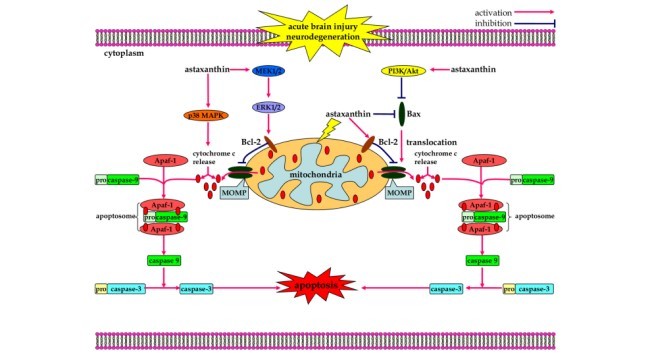
3.3 اثرات ضد آپاپتوزیس
4. نتیجه گیری و چشم اندازهای آتی
منابع
Abstract
1. Introduction
2. Astaxanthin: Source, Biochemistry, Bioavailability, and Safety
3. Neuroprotective Properties of Astaxanthin in Neurological Diseases
3.1. Anti-Oxidant Effects
3.2. Anti-Inflammatory Effects
3.3. Anti-Apoptotic Effects
4. Conclusions and Perspective
References